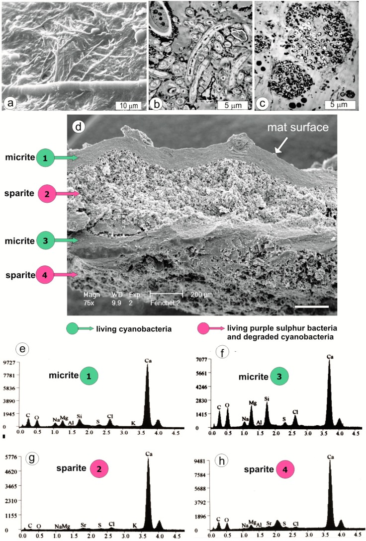
Figure 3.
(a) SEM image of the artificial mat surface (hot air-dried shrunken specimen) showing thinner filaments of Pseudanabaena and thicker filaments of Calothrix. (b) TEM image of Pseudanabaena and Calothrix filaments from about 0.3 mm below the mat surface. Note the large volume of extracellular polymers (EPS) excreted by the cyanobacterial filaments, which are incidentally associated with much smaller heterotrophic bacteria, and more rarely Mg calcite nanograins (opaque matter) precipitated in larger accumulations particularly on Calothrix filament (upper left corner). (c) TEM image of colonies of purple sulfur bacteria (possibly Thiocapsa) characteristic of the mat layers 2, 4, 6 and 8 (see diagram in Figure 4). (d) Vertical section of hot air-dried fragment of the artificial mat showing shrunken and weakly mineralized living cyanobacterial layers (micrite) alternating with strongly mineralized layers (sparite) composed of degraded cyanobacteria (mostly empty sheaths) and purple sulfur bacteria. (e, f) SEM-EDS spectra to show the negligible presence of Mg silicate in Mg calcite from the surficial cyanobacterial layer (micrite 1) and much higher in the Mg calcite from deeper located cyanobacterial layer (micrite 3).

