
Fig. 4A–B.
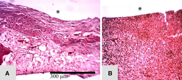
Histological features of the tissue response at 4 weeks postdébridement are seen in the following representative sections. (A) Healthy fibrous tissue is seen in the débridement site of a rabbit that received G-PNDJ (*débrided surface). (B) Thicker fibrous tissue invaded with acute inflammatory cells is seen in the débridement site of a rabbit that did not receive G-PNDJ. This reaction extends deep into the tissue, and there is new bone formation adjacent to the ulna (Stain, hematoxylin and eosin; original magnification, × 100) (*débrided surface).