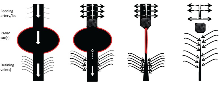
FIGURE 2.
Cartoon of usual behavior of macroscopic, discrete PAVMs pre and post embolization. (Left) Preferential blood flow through pulmonary arteriovenous malformation (AVM) sacs (red border) leads to reduced perfusion of non-PAVM associated arteries (gray), dilatation of feeding arteries that commonly appear as second or third order vessels; and dilatation/early filling of draining veins. Centre left: immediately following embolization of all feeding arteries, blood flow ceases through the pulmonary AVM, and is redirected to normal arteries. Surprisingly it appears very rare for sac thrombus to embolise before organization. Centre right and far right: over subsequent months, assuming feeding arteries remain occluded and the pulmonary AVM does not acquire new feeding arteries, organization, and remodeling leads to regression of the sac, and normalization of diameters of former feeding arteries/draining veins (For patient images, see Howard et al., 2014).

