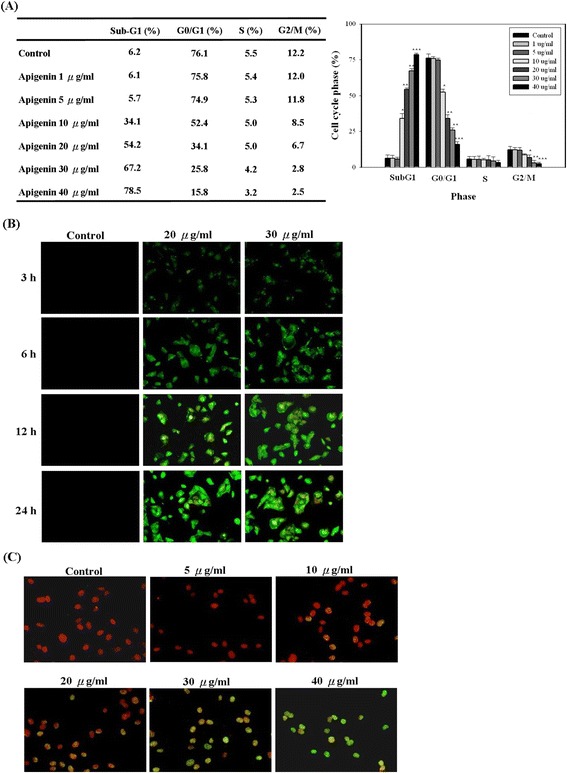
Figure 2.

Apigenin inhibited cell cycle progress and induced apoptosis in T24 cells. (A) Cultured cells were treated with or without apigenin under various concentrations (0–40 μg/ml). Twenty-four hours later, the cell cycle distribution was analysed by flow cytometry. The data indicate the percentage of cells in sub-G1, G0/G1, S, and G2/M phases of the cell cycle. (B) Cells were treated with apigenin (0, 20, and 30 μg/ml) for the indicated times, and then the induction of apoptosis was assessed by Annexin V-Alexa Fluor 488/PI assay kit. (C) Cells were treated with or without apigenin under various concentrations (0–40 μg/ml), and then the induction of apoptosis was assessed by TUNEL assay kit. The results represented the average of three independent experiments ± S.D. *P < 0.05, **P < 0.01, ***P < 0.001 compared with the untreated control.
