
Figure 3.
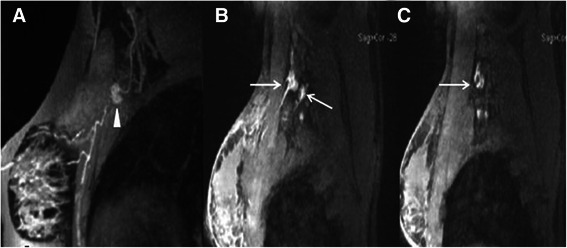
Comparison of MRL images between benign and malignant SLNs. A: Benign SLN in a 41-year-old woman with left breast ductal carcinoma. The lymph node displays homogeneous enhancement (white triangle) in Gd-MRL. B, C: Malignant SLNs in a 48-year-old woman with left breast ductal carcinoma. Heterogeneous enhancement and enhancement defect were found in Gd-MRL (white arrows).