Figure 2.

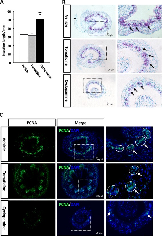
Inhibition of hedgehog signaling suppresses intestine shortening and remodeling at metamorphic climax. A: Cyclopamine-treated tadpoles have longer intestine at the climax. Tadpoles at stage 58 were treated with vehicle (100% ethanol, final concentration in rearing water: 0.1%) (9 tadpoles) or tomatidine (10 tadpoles), or cyclopamine (11 tadpoles) till they reached climax (stage 62). Intestine was isolated and measured from bile duct junction to colon. The animals in all three groups reached developed to stage 62 similarly as judged based on external morphology. ** indicate significant difference between the cyclopamine group and the two control groups (p < 0.001). B: Cyclopamine-treated tadpoles have fewer islets of adult epithelial stem cells. Transverse sections of the intestine from above were stained with methyl green pyronin Y (MGPY). The boxed area in the left panel is enlarged in the right panel. Note that at the climax of metamorphosis, the clusters (islets) of adult epithelial stem cells are stained strongly by MGPY (arrows) while the dying larval epithelial cells are only weakly stained [15,49,50]. The cyclopamine-treated tadpoles had smaller intestinal cross-section and fewer islets of adult cells. C: Cyclopamine-treated tadpoles have reduced cell proliferation. The intestinal sections above were stained with DAPI for nuclear DNA and anti-PCNA antibody for mitotic cells. The boxed area in the middle panel is enlarged in the right panel. Cyclopamine treatment significantly reduced cell proliferation in the intestine, especially in the epithelium. Note that the labeling in the epithelium was limited to the islet of the adult cells (arrows) but not in the dying larval epithelial cells, as expected.
