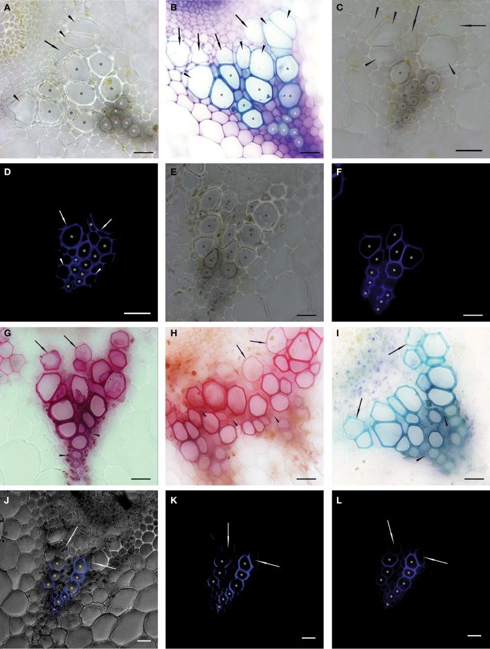
Figure 1.
Native cross sections of individual vascular bundles in inflorescence stems of Arabidopsis thaliana. Asterisks show vessels identified as functional (conductive) in 50 μm thick transverse sections observed in bright field microscopy—BF (A–C,E); epifluorescence microscopy—EF (D,F,L); and confocal scanning laser microscopy—CLSM (J,K). Vessels were identified as functional if their secondary cell wall was fully developed (BF) or more than one half of their perimeter was stained after proper setting of the exposure time (EF and CLSM). (A) Transverse section of non-stained vascular bundle. White asterisks mark protoxylem (PX) vessels and black asterisks mark metaxylem (MX) vessels identified as conductive. Arrowheads show expanded but not yet lignified MX vessels, arrows show expanding vessel cells (A–C). (B) Transverse section histochemically stained with toluidine blue prepared sequentially in a basipetal direction from the section in (A). White asterisks mark PX vessels and black asterisks mark fully lignified MX vessels considered as conductive. (C) Cross section prepared from apical stem segment perfused with Fluorescent Brightener 28 (FB28) dye solution. (D) The identical cross section to (C). Asterisks show stained secondary cell walls of conductive vessels perfused with the dye; non-lignified vessels are non-conductive and thus not stained; arrows show partially stained secondary cell walls of non-conductive MX vessels; arrowheads show stained fibers in PX area. (E) Cross section prepared from basal stem segment perfused with FB28 dye solution. (F) The identical cross section to (E). Compared to (E), seven fewer MX vessels were identified as conductive. (G–I) Images of vascular bundles in transverse sections prepared from basal segments perfused with basic fuchsine (G), safranin (H), and toluidine blue (I) solutions observed in BF. Arrows show stained but not yet lignified vessels; arrowheads show the intense staining of fibers. (J–L) An identical transverse section of a vascular bundle prepared from a basal segment perfused with FB28 dye solution and observed in CLSM (J,K) and in EF (L). Asterisks show the analogical number of vessels considered as conductive; arrows show partially stained vessels considered as non-conductive. (J) Permeation of a BF image with the signal from the FB28 solution (shown in K) perfused through conductive vessels and excited at a wavelength of 405 nm. Scale bars 20 μm.

