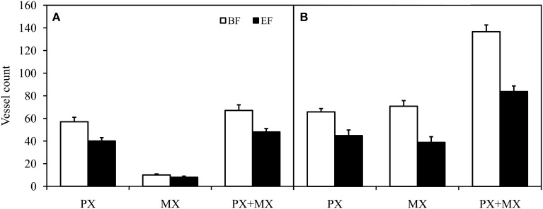
Figure 3.
Comparison of protoxylem (PX) conductive vessel count, metaxylem (MX) conductive vessel count, and total (PX + MX) conductive vessel count in apical (A) and basal (B) inflorescence stem segments of Arabidopsis thaliana. Conductive vessels were identified according to the development of secondary cell walls observed in bright field (BF) and the staining of secondary cell walls perfused with Fluorescent Brightener 28 dye solution and observed in epifluorescence (EF). There were statistically significant differences (P < 0.01) between conductive vessel identification methods in all groups of vessels. Means are given ± SE (n = 11).

