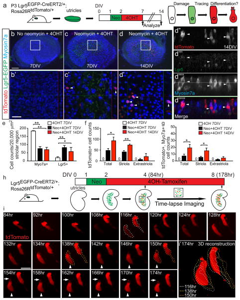
Figure 2. Lgr5+ cells act as striolar hair cell precursors in vitro.
(a) Utricles from P3 Lgr5EGFP-CreERT2/+; Rosa26RtdTomato/+ mice were treated with neomycin and then 4OH-tamoxifen (4OHT) to fate-map Lgr5+ cells. Organs were examined after 7 or 14 days in vitro (DIV). (b–b′) Undamaged controls had no tdTomato+ cells. (c–c′) Rare tdTomato+/Myo7a+ hair cells (HCs, arrowheads) were detected 7 days after damage, and (d–d′) many more double-labeled cells were found at 14DIV. (d″–d″‴) Orthogonal (XZ) views of d′. (e) Neomycin caused significant loss of striolar HCs and an increase in Lgr5+ cells. (f–g) Both total tdTomato+ cells and tdTomato+/Myo7a+ cells significantly increased over time. (h) Schematic for time-lapse imaging of lineage-traced Lgr5+ cells over 94 hr. (i) Stills of time-lapse (Supplementary Video 1) capturing two tdTomato+ cells selected from Lgr5-EGFP+ cells. tdTomato signal was first present in the right cell and later appeared in the left cell. Both cells first appeared tall and slender and resembled supporting cells. The left cell gradually became flask-shaped, beginning at 138 hr (arrowhead indicates hair cell-like cell). The basolateral portion of the left cell rounds up and an apical protrusion appeared around 154 hr (arrows). (i′) represents a 3D reconstruction of images at 174 hr. Dashed lines depict outlines of cells from stated time-points. n=3–6 in e–g. Data shown as mean±S.D. *p< 0.05, **p<0.01, Student’s t-tests. Scale bars, (b–d) 100 μm; (b′–d′) 20 μm; (i) 10 μm.

