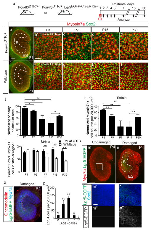
Figure 4. Lgr5+ supporting cells emerge after hair cell ablation in vivo.
(a) Schematic depicting the use of diphtheria toxin (DT) to ablate hair cells (HCs) in Pou4f3DTR/+ mice and Lgr5EGFP-CreERT2/+ mice to report Lgr5 expression. (b–e) DT treatment at P1 caused progressive HC loss and organ shrinkage over a 2-week period, followed by a partial recovery at P30. (f–i) Undamaged control organs from P3-P30. (j) Relative to age-matched, undamaged organs, DT-damaged organs were smaller. Damaged organs were the smallest at P15 and partly re-expanded at P30. (k) Normalized Myo7a+ cell counts similarly decreased and were the lowest at P15, before significantly increasing at P30 (p<0.0001, Student’s t-test). (l) Quantification showed that the damaged organs from Pou4f3-DTR mice contained higher percentages of Sox2+/Myo7a+ cells than age-matched, undamaged organs at P7, P15, and P30. (m) No detectable Lgr5-EGFP signals in undamaged organs at P4. (n) DT-mediated HC loss led to robust Lgr5-EGFP expression in striolar Sox2+ supporting cells (SCs) at P4 (also see Supplementary Fig. 3). 94.2±4.9% of Lgr5-EGFP+ SCs expressed Sox2+. (o) Lgr5-EGFP+ domain overlapped with the striola, where residual oncomodulin+ HCs resided. (p) Relative to age-matched, undamaged controls (Supplementary Fig. 3), there were significant more Lgr5-EGFP+ cells after HC ablation P2–7 (p<0.01, Student’s t-test). n=4–12 in j–l (4 for P3, 8 for P5, and 12 for P7, P15 and P30) and 4–6 in p. Data shown as mean±S.D. *p<0.05, **p<0.01, Student’s t-tests. Scale bars, (b, f, m, n, and o) 100 μm; (b′, c–e, f′, g–i, m′–n′, m″–n″) 20 μm.

