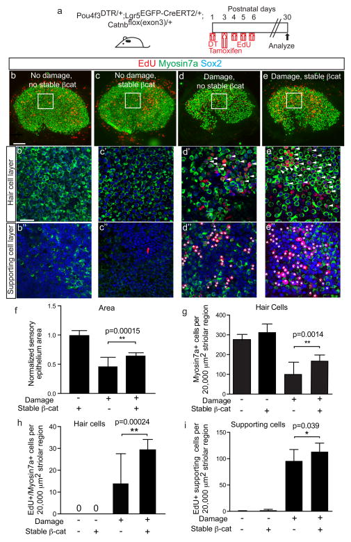
Figure 7. Stabilized β-catenin drives mitotic hair cell regeneration by Lgr5+ cells.
(a) Schematic of using Pou4f3DTR/+; Lgr5EGFP-CreERT2/+; Catnbflox(exon3)/+ mice to express stabilized β-catenin in Lgr5+ cells after hair cell ablation. EdU was administered to label proliferating cells. (b) Undamaged, controls showed almost no EdU labeling in the sensory epithelium. (c) Utricles from Lgr5EGFP-CreERT2/+; Catnbflox(exon3)/+ mice treated with tamoxifen also showed no increased EdU uptake. (d) Damage utricles showed fewer Myo7a+ hair cells (HCs) and increased EdU+ HCs and supporting cells (SCs) in the striolar region. (e) Stabilized β-catenin in Lgr5+ cells enhanced HC density and EdU labeling of HCs and SCs in the striolar region. (f–g) Sensory epithelium size and HC density significantly increased after β-catenin stabilization in Lgr5+ cells. (h–i) Relative to damage alone, β-catenin stabilization in Lgr5+ cells significantly increased the number of EdU+/Myo7a+ HCs and EdU+ SCs in the striolar region. n= 5–18 in f–i (18 for damaged organs without β-catenin stabilization, 5–8 for all other groups). Data shown as mean±S.D. *p<0.05, **p<0.01, Student’s t-tests. Scale bars, (b–e) 100 μm; (b′–e′, b″–e″) 20 μm.

