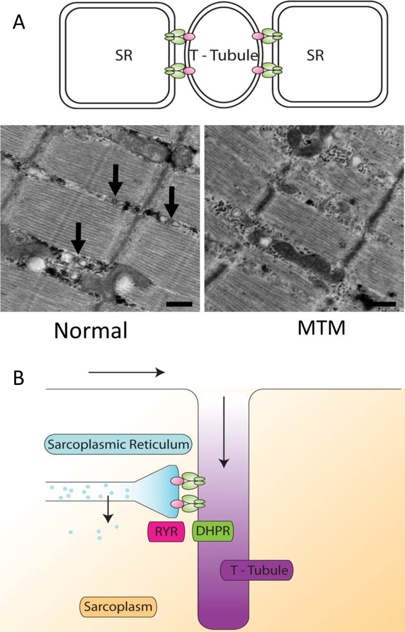
Fig. 1.
Structure and function of the triad. (A) The triad is a structure formed by the interface between the T-tubule and 2 portions of the sarcoplasmic reticulum (SR). It is normally seen on electron microscopy of longitudinal sections as a triplet of structures (arrows) between myofibrils and slightly offset from the Z-line. In triadopathies such as myotubular myopathy (MTM), the triad may be absent or disorganized. (B) The triad is a critical structure in the process of excitation–contractions coupling whereby electrical impulses (arrows) travel down in the membrane and into the T-tubules. Interaction between the dihydropyridine receptor (DHPR) in the T-tubule and the ryanodine receptor (RYR) in the SR produces the release of calcium from the SR into the sarcoplasm. This calcium then participates in a variety of cellular processes, including, especially, muscle contraction

