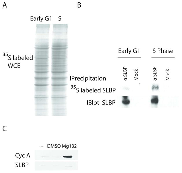
Figure 2. SLBP production rate is low in early G1 cells.
HeLa cells were synchronized by double thymidine block method and labeled with 35S-methionine for 15 minutes at early G1 (1.5–2 hours after M/G1) or S phase (1.5 hours after release) after half hour incubation in methionine lacking media. A) Equal protein amounts of total lysates and B) immunoprecipitates with SLBP antibody or beads alone were run on SDS PAGE gel and detected with autoradiography (top panel). Cells at the same cell cycle points were lysed and SLBP was immunoprecipitated from equal protein amounts of total lysates. Immunoprecipitated SLBP was detected by western blot using SLBP antibody (bottom panel) C) Early G1 cells similarly synchronized by double thymidine method were collected after one hour Mg132 (50 μM), DMSO (carrier) or just media treatment and western blot analysis was performed using Cyclin A or SLBP antibody as indicated. Cell cycle phases were confirmed by FACs analysis.

