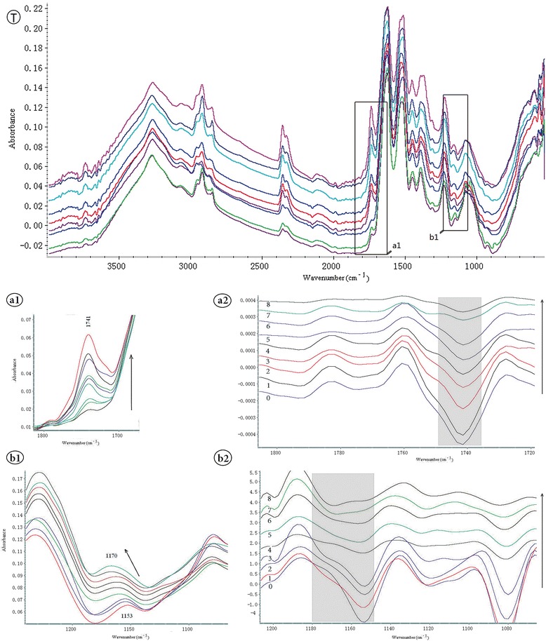
Figure 4.

ATR-FTIR spectra of MCF-7 cells treated with different concentrations of 5 -fluorouracil in Figure 4 T. From bottom to top correspond to drug concentrations including 0, 0.78, 1.56, 3.125, 6.25, 12.5, 25, 50 and 100 μg/mL. The increase in the band at 1741 cm−1 for cells exposed to increasing concentrations of 5-FU shows in a1 and the region at 1153–1170 cm−1 shows in b1. Corresponding secondary derivatives of the spectra are separately presented in a2 and b2 on the right.
