Figure 6.
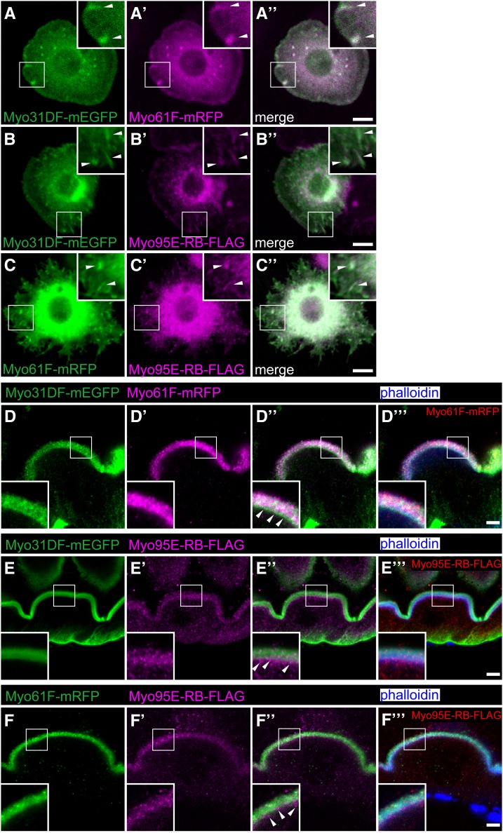
Colocalization of Drosophila class I myosin proteins. (A–C′′) S2 cells coexpressing Myo31DF-mEGFP (green) and Myo61F-mRFP (magenta) (A–A′′), Myo31DF-mEGFP (green) and Myo95E-RB-FLAG (magenta) (B–B′′), and Myo61F-mRFP (green) and Myo95E-RB-FLAG (magenta) (C–C′′). Insets in A–C′′ are high magnifications of the areas shown by white squares, and arrowheads in the insets indicate colocalization of the two coexpressed class I myosin proteins. A′′, B′′, and C′′ are merged images of A and A′, B and B′, and C and C′, respectively. (D–F′′′) The larval anterior midgut epithelial cells coexpressing Myo31DF-mEGFP (green) and Myo61F-mRFP (magenta) (D–D′′′), Myo31DF-mEGFP (green) and Myo95E-RB-FLAG (magenta) (E–E′′′), and Myo61F-mRFP (green) and Myo95E-RB-FLAG (magenta) (F–F′′′) were stained with fluorescently labeled phalloidin (blue). Insets in D–F′′′ are high magnifications of the areas shown by white squares. D′′, E′′, and F′′ are merged images of D and D′, E and E′, and F and F′, respectively. D′′′, E′′′, and F′′′ are merged images of fluorescently labeled phalloidin (blue) and D′′, E′′, and F′′, respectively. Bars, 5 μm.

