Figure 2.
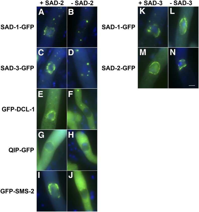
SAD-2 is required for the localization of other perinuclear MSUD proteins. (A–J) In the absence of SAD-2, other MSUD proteins lose their affinity for the nuclear periphery. (K–N) As with SAD-1 (Shiu et al. 2006), SAD-3 does not appear to affect the localization of others. GFP constructs were made according to Hammond et al. (2011b). (A) P13-14 × P13-15. (B) P21-37 × P21-38. (C) F4-30 × P14-59. (D) F6-31 × P21-32. (E) P21-39 × P21-40. (F) P21-30 × P21-31. (G) P15-62 × P15-63. (H) P21-26 × P21-27. (I) F5-06 × P15-14. (J) F6-30 × P21-28. (K) P13-14 × P13-15. (L) P21-35 × P21-36. (M) F2-23 × P21-41. (N) P21-33 × P21-34. Bar, 5 μm. [Note that a perinuclear MSUD protein first emerges as aggregates around the nuclei at the binuclear stage (Shiu et al. 2006). These aggregates coalesce after karyogamy and eventually become two opposite crescents during leptotene. The crescents spread into a ring-like structure during zygotene and pachytene. The ring becomes more patchy and irregular during the diffuse stage, and the perinuclear localization can no longer be seen after diplotene. A perinuclear MSUD protein may also appear as cytoplasmic foci between karyogamy and metaphase I. The exact nature of these foci is still unknown, although they have been speculated as some kind of endomembrane body.]

