Figure 2.

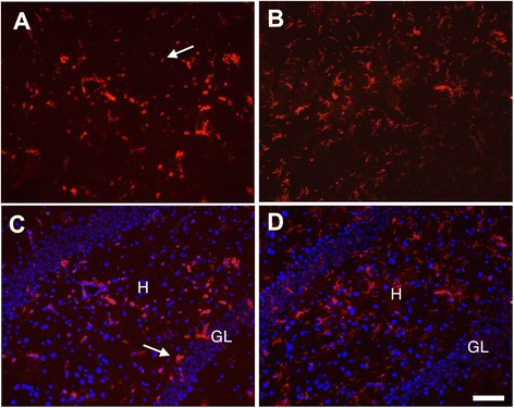
Neuregulin-1 inhibits DFP-induced changes in microglial morphology in the hippocampus. Brains collected 24 h post-DFP administration were immunostained for CD11b to identify microglia in the hippocampus, and a subset of these sections was also stained for DAPI (C, D). CD11b immunopositive cells with morphological characteristics of activated microglia (indicated by arrows) were seen in the hippocampus 24 h following DFP administration (A, C). In contrast, CD11b immunopositive cells in the hippocampus of DFP intoxicated animals treated with NRG-1 exhibited morphologies characteristic of resting microglia (B, D). H, hilus; GL, granule cell layer. Stereotaxic coordinate = bregma −3; Scale bar = 50 μm.
