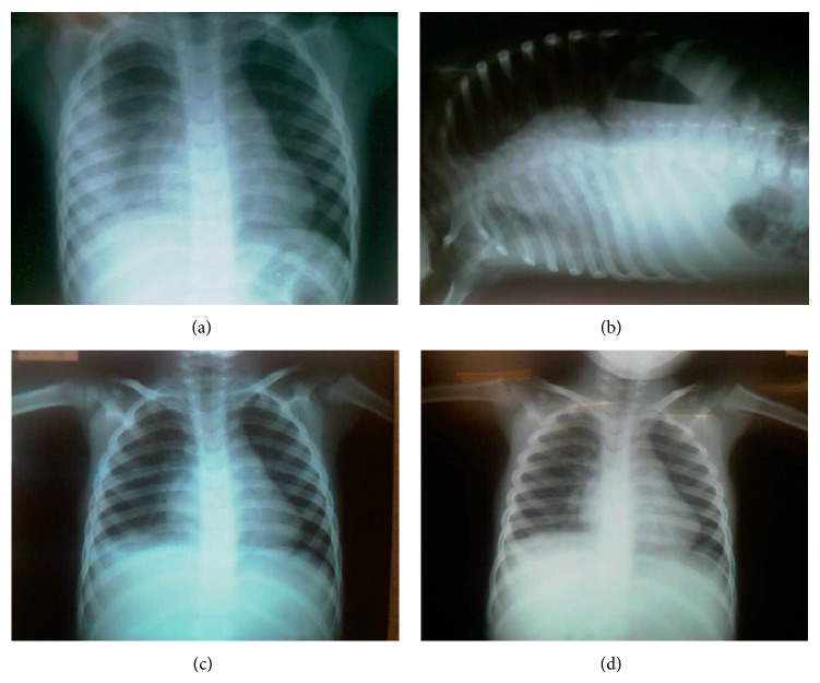
Figure 1.
(a) Chest radiograph on the anteroposterior incidence, on day 1 of hospitalization and 15th day of clinical symptoms of pneumonia, showing the lobar pneumonia and a beginning of atelectasis in the right hemithorax. (b) Chest radiography on the incidence of Laurell on 1st day of admission showing pleural effusion in the right hemithorax. (c) Chest radiography on the posterior-anterior incidence on the 4th day of hospitalization, showing atelectasis improvement. (d) Chest radiography on the posterior-anterior incidence on the last day of hospitalization showing atelectasis reversal.

