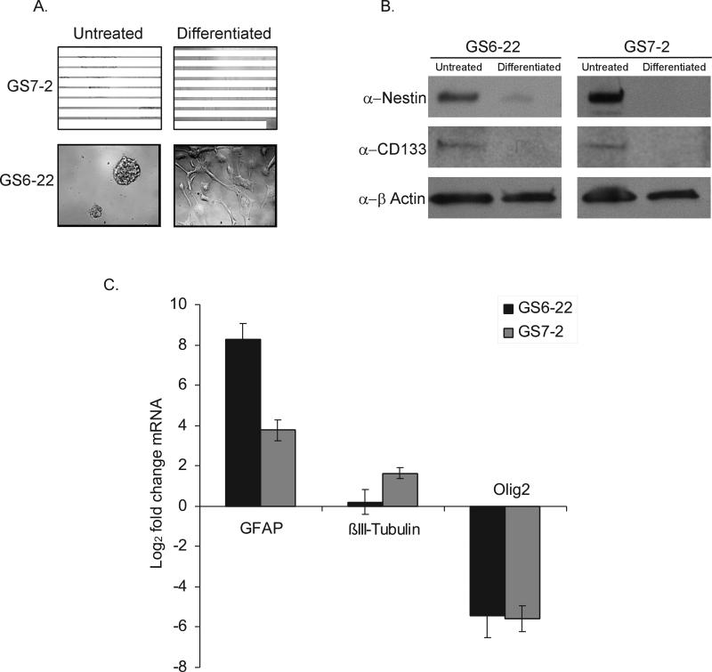
Figure 1. Glioblastoma stem cells derived from primary tumor samples form neurospheres in culture, express neural stem cell markers, and retain the ability to differentiate.
A. Images of GS7-2 and GS6-22 neurospheres grown in serum-free media with EGF and FGF-2 (Untreated) or differentiated by plating on poly-L-ornithine and laminin in the presence of 2% serum without growth factors (Differentiated). B. Immunoblot of lysates of GS7-2 and GS-22 neurospheres in serum-free media or after 7 days under differentiation conditions with antibodies to CD133, nestin, and β-actin. C. RT-qPCR analysis of GFAP, βIII-tubulin, and olig2 mRNA in GS7-2 and GS6-22 neurospheres exposed to differentiation conditions for 7 days. Fold change values were calculated relative to untreated control and normalized by comparison to β-actin and plotted as log2. Bars represent mean of three separate experiments; bars SD.

