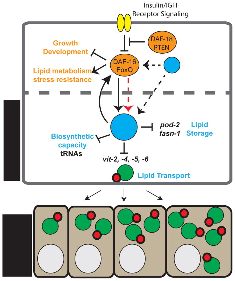
Figure 6. Model of the central role of MAFR-1 in organismal physiology.
MAFR-1 can influence animal physiology in both an insulin/FoxO-dependent (orange) and independent (blue) manner. Altered mafr-1 levels in the intestine cell non-autonomously impacts fecundity through changes in the production of the vitellogenin family of proteins that deregulates lipid transport to developing oocytes in the germ line. Black arrows and bars indicate genetic interactions. DAF-16/FoxO may directly regulate the expression of mafr-1 (dashed red arrow). Other factors that regulate DAF-16/FoxO-dependent and independent regulation of Maf1 may exist (?) and influence Maf1 cell autonomously and non-autonomously. Dashed cell boundary indicates potential auto-, para-, or endocrine signals resulting from Maf1 activity.

