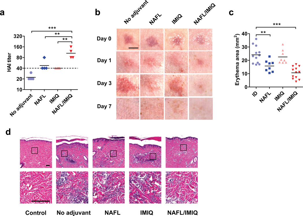
Figure 3. NAFL/IMIQ adjuvant in swine study.
The exterior hind legs of yorkshire pigs were shaved, cleaned and ID vaccinated with 100 µl of the influenza vaccine (3 µg HA content) only (no adjuvant) or following one pass of laser illumination with Fraxel SR-1500 (NAFL), after which IMIQ was applied to the immunization site (NAFL/IMIQ). Alternatively, the immunization site receiving the vaccine alone was topically applied with IMIQ directly (IMIQ). HAI titers were measured in 2 weeks (a). Each symbol represents data from individual animals, horizontal bars indicate mean, and a dish line marks the standard protective titer of HAI. n=4. (b) Photos were taken right (day 0) and 1, 3, and 7 days after immunization and representative results were shown with 4 pigs in each group. Scale bar, 5 mm. (c) The erythema areas of injection sites were analyzed by Image Pro Premier software 3 days after immunization. Each symbol represents a mean value of one injection site analyzed for 3 times. Horizontal bars indicate the mean. From left to right, n=11, 8, 8, and 12, respectively. (d) H&E slides showing infiltrated cells at the inoculation site, representative of 6 similar results in two separate experiments. Scale bar, 200 µm. Statistical significance was analyzed by ANOVA/Bonferroni. *, p<0.05; **, p< 0.01 or ***, p<0.001, respectively.

