Figure 1.

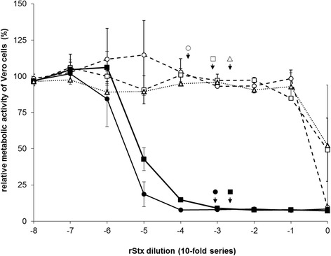
Effect of recombinant Shiga toxins and toxoids on the cellular metabolic activity of Vero cells. Cells were incubated for 96 h at 37 °C with 10-fold dilutions of endotoxin-deprived lysates prepared from E. coli BLR(DE3) transformed with plasmids encoding for rStx1WT (filled circle, solid line), rStx1mut (open circle, dashed line), rStx2WT (filled square, solid line), rStx2mut (open square, dashed line) or vector control (open triangle, dashed line). Results of VCA are presented relative to data obtained with cells incubated with plain medium as negative control (set to 100%) and data from cells treated with 1% SDS as positive control (set to 0%). Data is depicted as means ± standard deviations from duplicate determinations in one representative out of four independent experiments. Missing error bars are within symbols. For functional assays with bovine primary cell cultures, lysates containing rStxWT were adjusted to reach a final concentration of 200 verocytotoxic doses 50% per mL. Lysates containing rStxmut were diluted to yield the same OD as the corresponding rStxWT–containing lysate in an ELISA assay (for details see Material and methods). To visualize the verocytotoxic activities of the respective rStx working dilutions, the calculated dilution factors are depicted by arrows and a corresponding symbol in the diagram.
