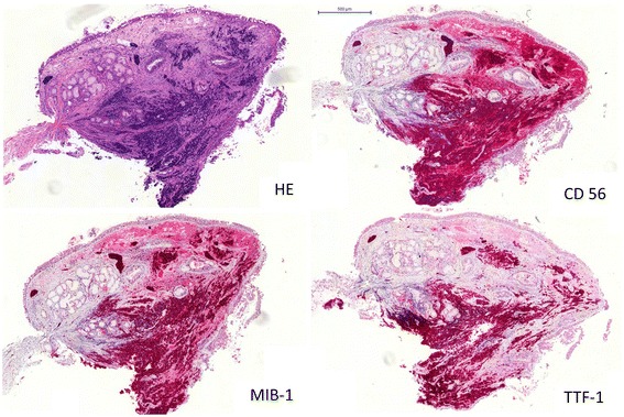
Figure 2.

Example of TTF-1 positive small cell lung cancer (SCLC) staining. The tumor was identified as TTF-1 positive when more than 5% of cells stained for TTF-1. Left upper panel: Hematoxylin and eosin stain (HE). Right upper panel: Neural cell adhesion molecule (CD56). Left lower panel: Ki-67 Proliferation marker (Clone MIB-1). Right lower panel: Thyroid transcription factor-1 (TTF-1).
