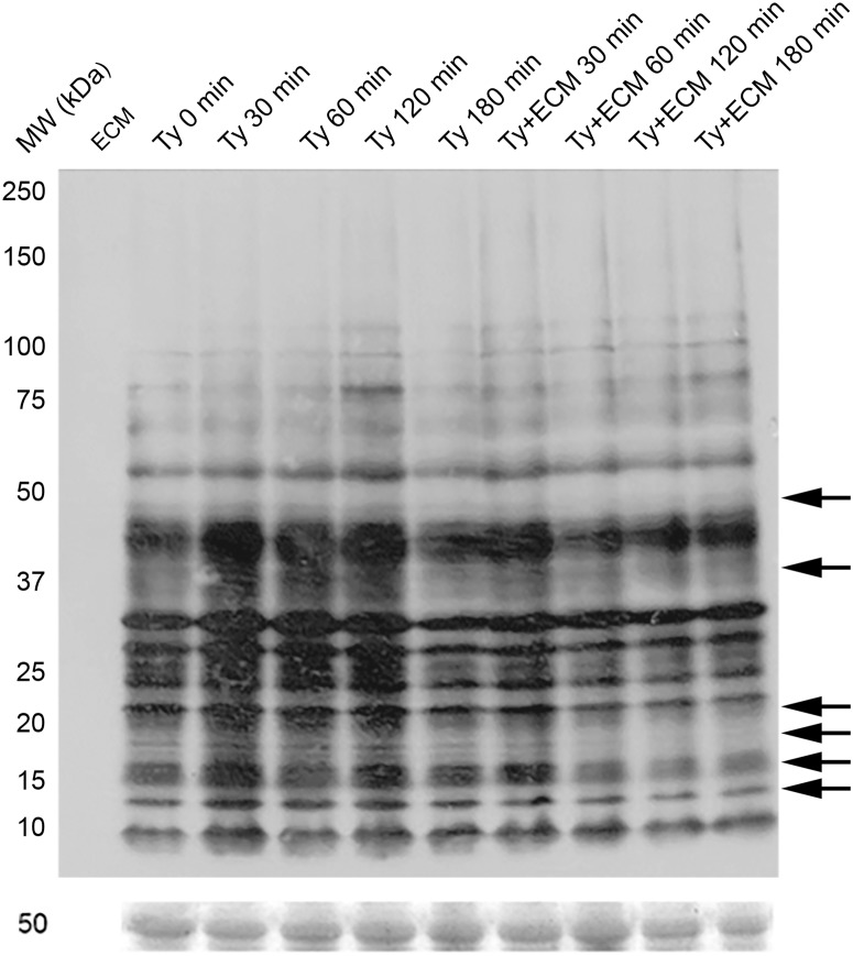
Fig 3. S-nitrosylation pattern during Trypanosoma cruzi trypomastigotes adhesion to extracellular matrix.
Trypomastigotes (1x10⁹) were incubated with ECM (1.5 mg) in phenol red free-MEM, supplemented with 2% FBS, at 37°C and 5% CO2, for different incubation times. Proteins (50 μg) were resolved in 6–16% gradient SDS-PAGE. The immunoblotting was performed using the antibodies: rabbit anti-SNO 1:2,000, and the secondary HRP conjugated anti-rabbit 1:8,000. The Ponceau’s staining was used as a load control. Arrows indicate protein bands in which the signal intensity is decreased on the group incubated with the extracellular matrix in relation to the equivalent group incubated with growth media alone. The figure is representative of three experiments.

