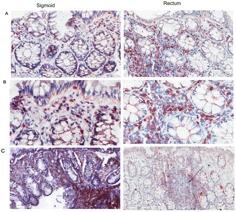
Fig 1. CD3+ lymphocytes are similar in normal human sigmoid colonic and rectal mucosa.
Sigmoid colonic and rectal mucosal sections immunostained for CD3. The membrane red/brown colors denote positive staining (red/brown, AEC chromogen). Specimens were counterstained with hematoxylin. (A)Upper panel original magnification 200x (B) lower panel 400x (C) lower magnification showing distribution of lymphoid aggregates in both sigmoid and rectum (100x). Arrows represent lymphoid aggregates.

