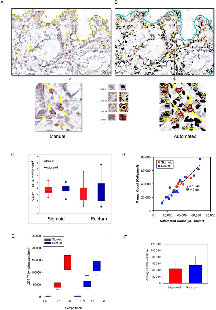
Fig 2. Application of an automated algorithm to quantitate T lymphocytes in immunostained sigmoid colonic and rectal mucosa.
A membrane detection algorithm was applied to CD3-immunostained sigmoid colonic and rectal mucosal biopsies from 5 healthy donors (5 sigmoid colon, 5 rectum). (A) Digital images with AEC chromogen staining for CD3 and hematoxylin counterstaining. (B) Mark-up images with red, orange, and yellow pixels depicting immunopositive cells (strong, moderate, and weak intensity, respectively), and black pixels depicting nuclei for immunonegative cells. Bar = 100 μm. Arrowheads denote manual enumeration. (C) Tissue concentrations of T lymphocytes obtained using both methods and analyzed by paired t test. (D) Correlation between counts obtained by manual and automated enumeration of the same regions. R2 value = 0.90 (E) Tissue concentrations of T lymphocytes in epidermis (Epi), lamina propria (Lp), and lymphoid aggregates (La; when present) of sigmoid colon and rectum. (F) Average T lymphocyte concentrations obtained from averaging the various compartments from the sigmoid colon and rectum.

