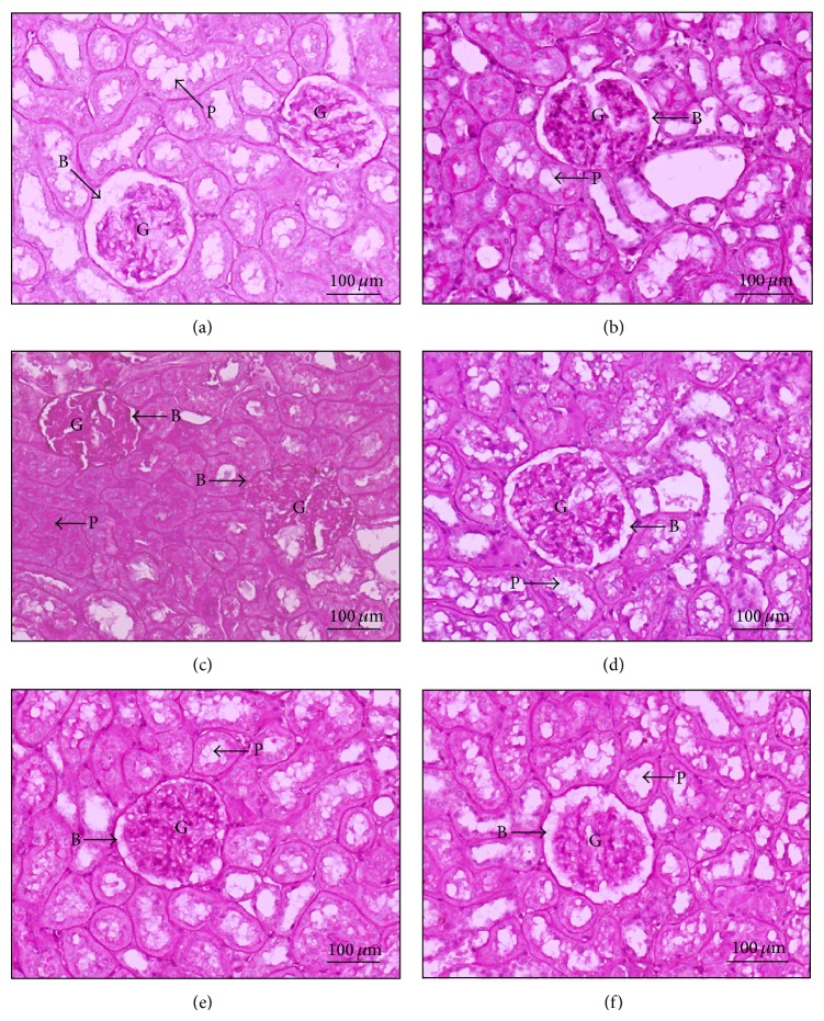
Figure 3.
Micrographs of Periodic Acid-Schiff (PAS) staining of rat kidneys. Light microscopies of sagittal half of kidney sections stained with PAS and counterstained with hematoxylin are shown (original magnification 200x for all panels). (a) Normal (ND), (b) normal diet supplemented with CGE (ND + CGE), (c) type 2 diabetes mellitus (DM), and (d) T2DM supplemented with CGE (DM + CGE). Bowman's capsular spaces are indicated by arrow “B”; the proximal tubular epithelium and the lumen are indicated by arrow “P.” Glomerulus (G).

