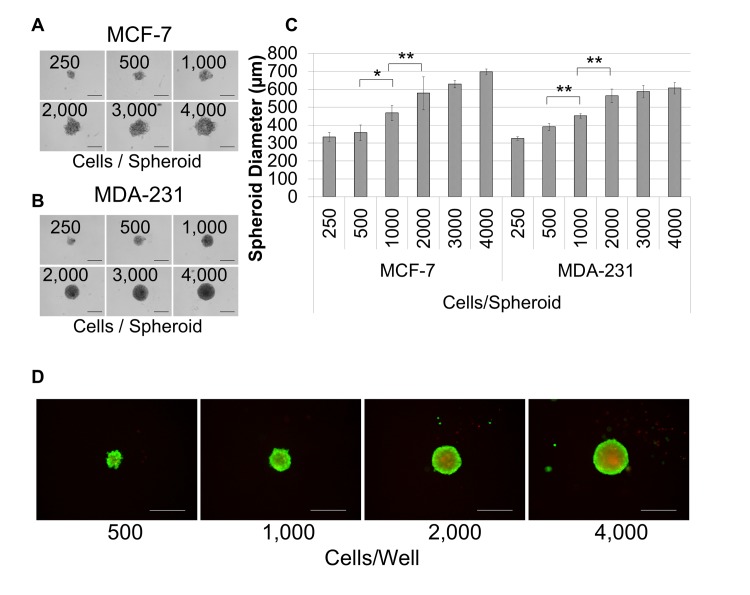
Fig 1. The optimal breast cancer cell seeding density for spheroid formation is 2,000 cells/well.
Breast cancer cells were seeded at the concentrations (cells/well) shown in 96 well, ultra-low adhesion plates and incubated for 72 hours in 1X spheroid formation ECM for MCF-7 (A) and MDA-MB-231 (B) cells. C. The diameter of each spheroid was measured for quadruplicate samples and groups were compared for statistical significance using the student’s t-test for both cell lines. D. Non-fluorescent calcein am was converted to fluorescent calcein (green), indicating living cells, and ethidium bromide (red) was internalized by dead cells. The presence of live cells in the outer layers of the spheroid and dead cells in the spheroid core is indicative of physiological diffusion gradients. Scale bar = 500 μm. *P < 0.05, **P < 0.01.

