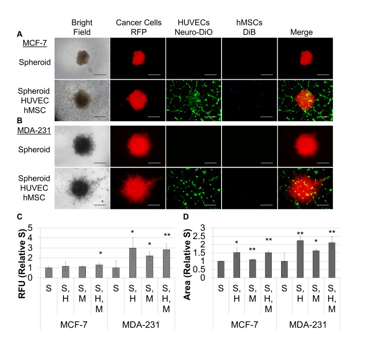
Fig 3. hMSCs and endothelial tubules promote invasion and differentially effect proliferation of MDA-MB-231 and MCF-7 spheroids.
A. Spheroids formed as described above under hypoxia. Then 96 well, flat bottom plates were coated with 50 μl of tubule formation matrix and incubated for one hour to polymerize the hydrogel. For wells with HUVECs, 12,500 cells were added to each well, and for remaining samples, EGM-2 was added. HUVECs were allowed to assemble into tubules for two hours. One spheroid was transferred to each of the wells in the plate. MCTS were allowed to settle for 1 hour. Then, 100 μl of medium was aspirated from each well. For wells with hMSCs, 10,000 cells were suspended in each ml of Invasion Matrix, and 50 μl was added to each well. For the remaining samples, 50 μl of tumor-aligned invasion matrix was added to each well. The plates were then incubated at 37°C, 5% CO2 for 1 hour to polymerize the hydrogel, and 100 μl of TARPMI, 10% FBS was added to each well. Cultures were incubated under hypoxia for 96 hours. Images are provided for spheroids alone and for spheroids, HUVECs, and hMSCs for MCF-7 (A) and MDA-231 (B). Cultures were analyzed as described above for proliferation (C) and invasion (D), and samples were evaluated in quadruplicate. S = breast cancer MCTS, H = HUVEC network, and M = hMSCs. Scale bar = 500 μm. *P < 0.05, **P < 0.01.

