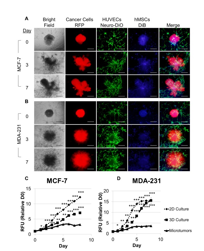
Fig 4. Microtumors of MCTS, hMSC, and endothelial tubules produce a physiological breast cancer niche possessing tumor morphology, tumor invasion, and endothelial recruitment and exhibiting differential cell proliferation compared to 2D and 3D monocultures.
Microtumors were modified from Fig 3 such that hMSCs at 1,000 cells/well were added first to the HUVECs that had been incubated for 2 hours. After one hour of HUVEC/hMSC coculture, one MCF-7 spheroid (A) or MDA-MB-231 spheroid (B) containing 500 cells/well hMSCs was added to each well, embedded in TA Invasion Matrix, and overlaid with TARPMI, 10% FBS. A direct comparison of cell proliferation was made between microtumors, 2D monoculture, and 3D monoculture based on fluorescence intensity of the RPF-expressing MCF-7 (C) and MDA-MB-231 (D) cells over eight days in culture. Values were normalized to fluorescence intensity of the onset of assay for each cell line and culture condition, and samples were evaluated in quadruplicate. Scale bar = 500 μm. *P < 0.05, **P < 0.01, ***P < 0.001.

