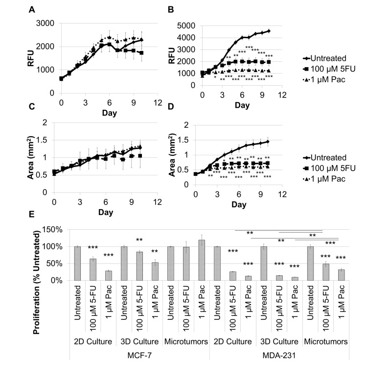
Fig 6. Microtumors exhibit physiological responses to paclitaxel or fluorouracil treatment, and microtumors exhibit a decrease in breast cancer cell proliferation compared to 2D and 3D monocultures for MCF-7 and MDA-MB-231 cells after 7 days of treatment.
Microtumor growth and response to 100 μM fluorouracil or 1 μM paclitaxel were determined based on fluorescence intensity of the RPF-expressing MCF-7 (A) and MDA-MB-231 (B) cells over ten days. A similar response was observed for the increase in microtumor area as an indicator of spread or invasion for these treatments for the MCF-7 (C) and MDA-MB-231 (D) microtumors. E. A direct comparison was made between 2D culture, 3D culture, and microtumors on day 7 for MCF-7 and MDA-MB-231 in the presence of fluorouracil or paclitaxel. Fluorouracil and paclitaxel inhibit breast cancer cell proliferation and spread for MDA-MB-231 microtumors, but not for MCF-7 microtumors. In addition, 2D and 3D monocultures were hypersensitive to fluorouracil or paclitaxel treatment compared to microtumor for both cell lines. Samples were evaluated in quadruplicate. *P < 0.05, **P < 0.01, ***P < 0.001.

