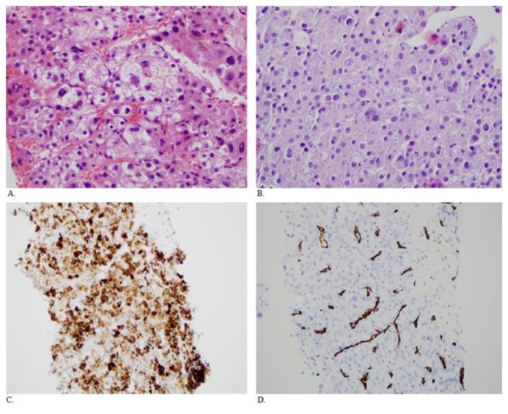
Figure 2.
Fifty year old male with HCC. A. Sections of the original liver needle core biopsies show a moderately differentiated hepatocellular carcinoma with a trabecular growth pattern. There are focally bizarre nuclei. (H&E, 400× original magnification) B. Sections of the rectus mass core biopsy show similar morphology to the original biopsy taken over 5 years previously (H&E, 400× original maginification). C. Immunohistochemistry shows the tumor cells are positive for the hepatocyte marker Hep Par 1 (clone OCH1E5) (200× original magnification). D. There is also CD34 immunopositivity in the endothelial cells, typical of hepatocellular carcinoma (200× original magnification). The morphology and the stains support the diagnosis of a metastatic hepatocellular carcinoma.

