Figure 3.
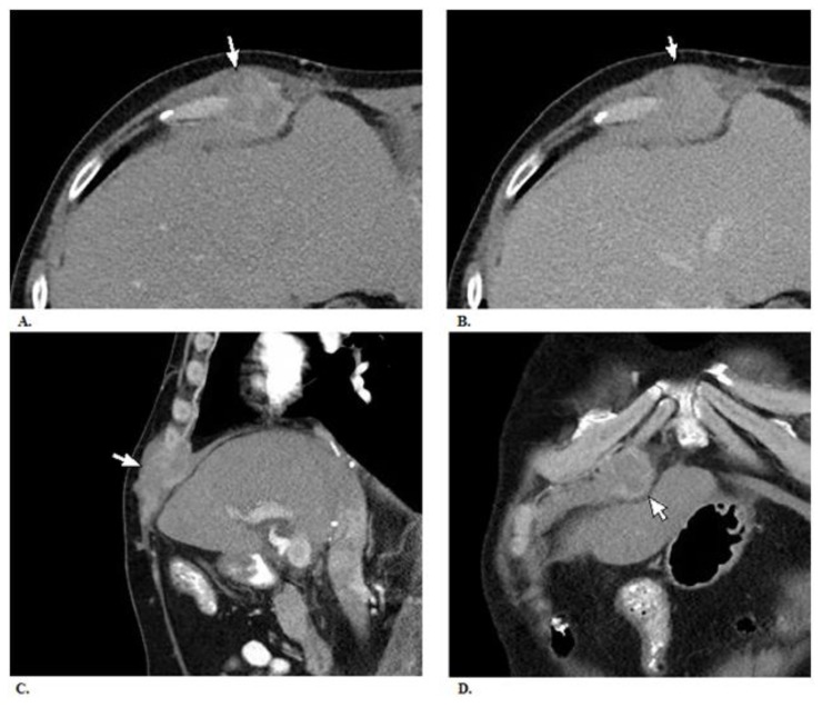
Fifty five year old male with biopsy needle tract seeding from HCC. A. There is a mass measuring 4.3 × 3.8 × 6.1 cm within the right rectus sheath (arrow). The mass demonstrates heterogeneous enhancement on venous phase images. B. Its enhancement is less intense and similar to that of the liver on delayed phase images. C. & D. Sagittal and coronal reconstructions of venous phase images showing the mass. It appears to invade the adjacent costochondral cartilage. Mild thickening of the anterior right hemidiaphragm suggests possible involvement. No obvious invasion of the underlying liver is seen, when accoutning for volume averaging artifact. There is no evidence of spread into the subcutaneous fat. TECHNIQUE: Contrast enhanced CT (120 cc iohexol) CT images in venous phase (100 kVp, 295 mAs, 5mm slice thickness, scan obtained 60 seconds following injection of contrast) and delayed phase (100 kVp, 261 mAs, 5mm slice thickness, scan obtained 3 minutes following injection of contrast) obtained on Siemens SOMATOM Definition AS.

