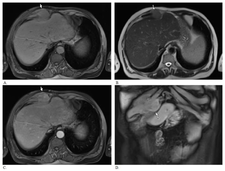
Figure 4.
MRI of 55 year old male with needle tract seeding from HCC 5 years following percutaneous biopsy. MRI demonstrates right rectus sheath enhancing mass (arrow) abutting and possibly invading the adjascent costochondral cartilage. There is no obvious invasion of the adjacent liver. A. Axial T1 weighted Volumetric Interpolated Breath Hold Examination image (VIBE, TR 7.22, TE 3.38, 4mm slice thickness, 1.5 Tesla). B. Axial T2 weighted Half-Fourier Acquisition Single-Shot Turbo Spin-Echo image (HASTE, TR 1000, TE 80, 6mm slice thickness, 1.5 Tesla). C. & D. Axial and coronal post gadolinium T1 weighted images (VIBE, arterial phase after administration of 19 cc gadodiamide intravenously, TR 7.22, TE 3.38, 4 mm slice thickness).

