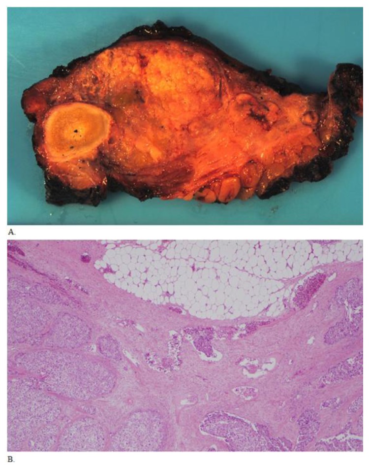
Figure 5.
Fifty five year old male with biopsy needle tract seeding by HCC, status post resection of the metastatic mass. A. Grossly, the abdominal wall mass resection specimen had tan-pink lobular cut surfaces with no bony invasion. B. Histologic sections show similar morphology compared to the two previous biopsies. Extensive lymphovascular invasion is present, and is seen in the center of the field (40× original magnification).

