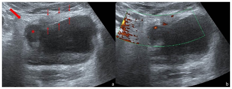
Figure 1.
Thirty-one year old woman with inflammatory pseudotumor of the urinary bladder. Abdominal US scan performed with a convex array transducer (3–6 MHz) on a Toshiba Aplio 500®. Axial scanning plane through the hypogastrium (a) demonstrates the partially filled urinary bladder, with a thickened wall (thin arrows). Maximum thickness was 21mm. In the right-anterior segment of the thickened wall there is a round, hypoechoic area (*) of 19mm diameter. The surrounding fat is hyperechoic (thick arrow). Power Doppler imaging in the same axial scanning plane (b) shows vascularization within the thickened wall.

