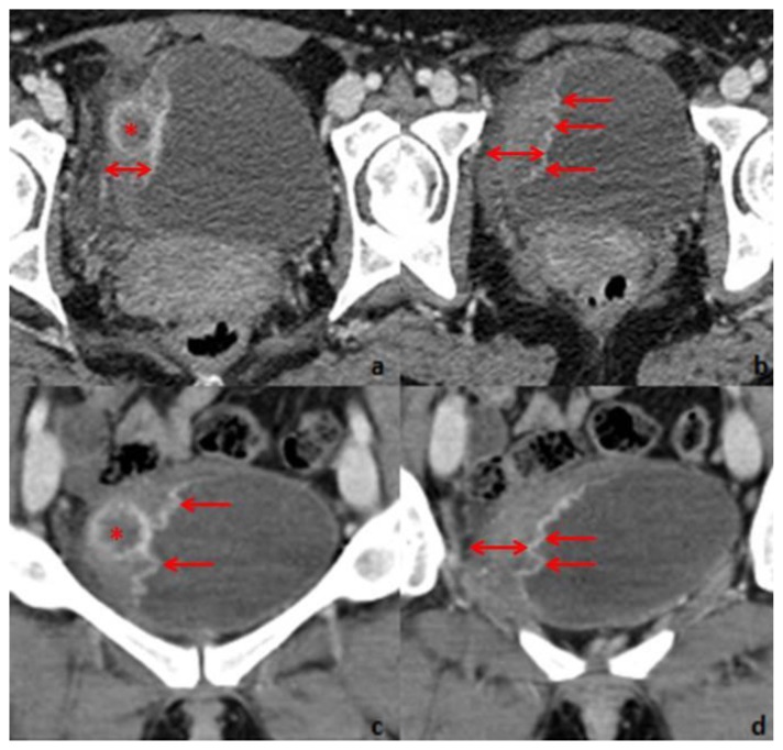
Figure 2.
Thirty-one year old woman with inflammatory pseudotumor of the urinary bladder. Contrast-enhanced CT, performed with a SimensSensation® (64 slice-CT). Radiation dose: 120Kv and 70 mAs. Injection of 120ml of 400mg/ml iodine concentration non-ionic contrast agent (Iomeron®). Images selected were taken 60s after contrast injection. Axial 1,5 thickness images (a and b) and coronal 4mm thickness reconstruction images (c and d) demonstrate right lateral bladder wall thickening (double arrow). Maximum thickness is 21mm. The mucosal surface is rim enhancing and has multiple papillary projections protruding into the urinary bladder lumen. Within the thickened wall, there is a nodular, 20mm diameter hypoenhancing area (*) with hyperenhancing margins.

