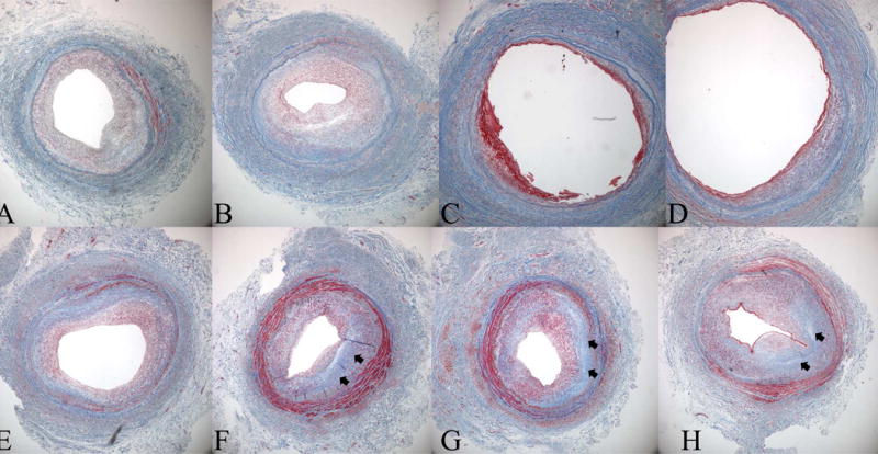
Figure 2.

Histological evidence for negative remodeling and intimal hyperplasia as a cause of late lumen loss in a great saphenous vein bypass graft. Masson's Trichrome sections are from an 8 month old femoro-posterior tibial artery vein bypass graft that was explantated due to hemodynamically significant stenosis identified by surveillance duplex ultrasound. The repair was constructed by an interposition graft and an 8 cm piece of diseased segment was explanted, registered, and serially sectioned from A (proximal graft) to H (distal graft). Note two areas of significant stenosis, sections A and B, and sections F-H with an intervening area of relatively normal vein. While the vein was uniform size and luminal caliber at the time of original surgery the stenotic areas demonstrate loss of total vessel area indicating lumen loss is due not only to initimal hyperplasia but also negative remodeling. Note the amount of fibrous protein, blue stain (arrows), in the stenotic segments of the graft.
