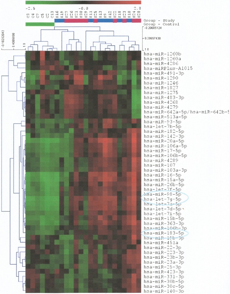
Fig 2. Heat map diagram showing the expression of the 50 miRNAs with the highest standard deviation in all samples.
The color scale illustrates the relative expression level of miRNAs and specifically, red color represents an expression level below the reference channel, whereas green color represents an expression higher than the reference. Each row represents a microRNA and each column represents a sample. The microRNA clustering tree is shown on the left. The control and study groups are clearly indicated with different colours. Samples S9-S13, S16, S18 & S19 correspond to the study group of patients with schizophrenia and tumor formation, whereas samples S21-S30 correspond to the study group of patients with tumor formation only. The 3 miRNAs, the expression of which was found to be significantly higher in the samples of the control group of patients, are also indicated.

