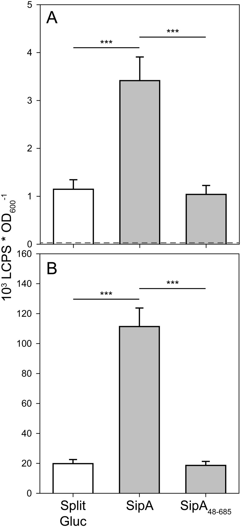
Fig 5. Gateway-based split Gluc protein complementation assay (PCA) for detection of chaperone-effector interaction.
Expression clones encoding InvB fused to GlucM43L 105 and SipA or a SipA variant lacking its CDB (SipA48–685) fused to GlucM110L 106 or GlucM43L 105 and GlucM110L 106 alone (split Gluc) were transferred into S. Typhimurium and expression was induced with 50 ng*ml-1 AHT. Gluc activity was quantified from intact cells (A) and bacterial lysates (B). The dotted line in (A) represents the background luminescence determined with an empty vector control (pWSK29). Mean and standard deviation out of three independent experiments done in triplicate is shown. Statistical analysis by Student’s t-test was done by comparing individual strains as depicted: ***, P < 0.001.

