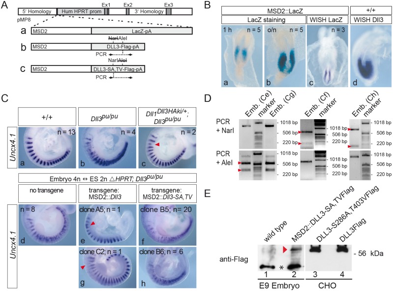
Fig 5. The DLL3-S286A,T403V mutant does not complement the loss of endogenous DLL3 in somitogenesis.
(A) Schematic representation of constructs introduced into HPRT deficient homozygous Dll3 mutant ES cells. MSD2 is a dimer of the mesoderm-specific promoter element (MSD) from the Dll1 locus [59]. Locations of primers and restriction sites used for genotyping of embryos derived from tetraploid complementation are indicated below and above schemes b and c, respectively. (B) The MSD2 promoter element drives transgene expression in the PSM similar to endogenous Dll3 expression. E8.5 MSD2::LacZ chimeric embryos were stained for β-galactosidase activity for one hour (a) and over night (b), or were analyzed by whole-mount in situ hybridization with a lacZ specific probe (c). (d) Expression pattern of endogenous Dll3 transcripts in an E8.5 wt embryo for comparison. (C) Whole mount in situ hybridizations of E9.5 wt (a), homozygous Dll3 pu (b), and Dll1 Dll3ki, Dll3 pu/pu [38] (c) embryos. In Dll1 Dll3ki, Dll3 pu/pu embryos, which lack endogenous DLL3 but express DLL3 from the Dll1 locus, expression of the anterior-posterior (A-P) somite patterning marker Uncx4.1 is restored except for minor irregularities (arrowhead in c). (d-h) Completely ES cell-derived embryos hybridized with Uncx4.1. Embryos generated from ES cells homozygous mutant for Dll3 (Dll3 pu/pu) and carrying the HPRT deletion (ΔHPRT) display the same A-P somite patterning defects as homozygous Dll3 pu embryos (compare d with b). Expression of wild type DLL3 in ES cell-derived embryos almost completely rescues the Dll3 pu A-P somite patterning defect except for minor irregularities (arrowheads in e, g), whereas ES cell-derived embryos expressing mutant DLL3 display a pudgy-like somite phenotype (f, h) indicating that DLL3-S286A, T403V is not functional during somitogenesis. (D) Genotyping of tetraploid embryos shown in (C) using PCR and restriction digests as indicated in (A). The wild type Dll3 PCR product is cut by AleI but not NarI (left panel), whereas the Dll3-S286A, T403V PCR product is cut by NarI (due to the presence of S286A) but not by AleI (due to the presence of T403V; middle and right panel; see Material and Methods for further details). Letters in parentheses refer to embryos shown in (C), arrowheads indicate cleavage products of the expected sizes. (E) Western blot analysis of lysates of wild type embryos (lane 1) or embryos obtained with ES cell clone B5 (lane 2) and lysates of CHO cells overexpressing Flag-tagged DLL3-S286A, T403V (lane 3) or wild type DLL3 (lane 4) using anti-Flag antibodies confirmed expression of DLL3-S286A, T403V-Flag in completely ES cell-derived embryos (red arrowhead). The equivalent of 4 trunks of E9 embryos was loaded in lanes 1 and 2. Asterisk between lanes 1 and 2 indicates a background band detected in embryo lysates.

