Figure 1.

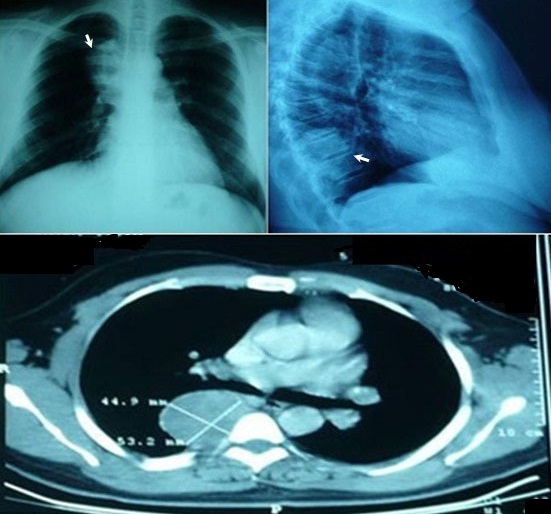
Chest x-ray (top) and computed tomography (bottom) revealing a posterior and well-circumscribed paravertebral mass, corresponding to a schwannoma

Chest x-ray (top) and computed tomography (bottom) revealing a posterior and well-circumscribed paravertebral mass, corresponding to a schwannoma