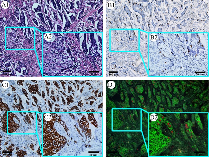
Fig 3. Comparison between QDs-based multiple imaging and traditional staining.
HE staining can show histological structures, but not label Ki67 and tumor nests well (A1 and A2). Immunohistochemical staining on Ki67 only can label Ki67 clearly, but not on tumor nests well (B1 and B2). Immunohistochemical staining on CK can label tumor nests clearly, but not on Ki67 well (B1 and B2). QDs-based multiple imaging can simultaneously label Ki67 and tumor nests clearly (D1 and D2). (In each panel, image 2 is the magnification of the square part of image 1. Magnification: A1, B1, C1 and D1: 100×; A2, B2, C2 and D2: 200×). QDs: quantum dots; HE: hematoxylin and eosin; CK: cytokeratin.

