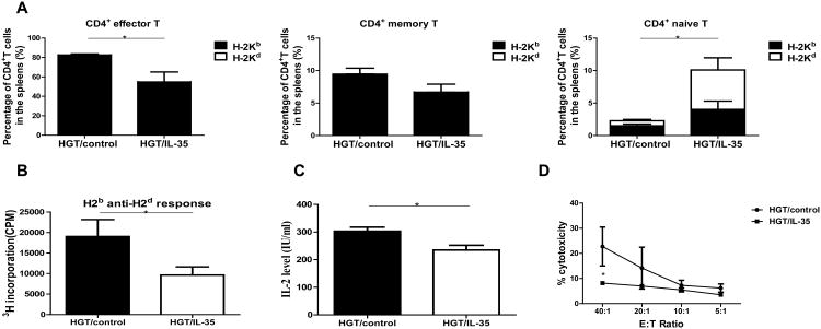
Figure 2. IL-35 led to a reduction of CD4+ effector T cells and reduced alloreactivity.
The experiments were performed as described in Figure 1. Lymphocytes were isolated from spleen, liver, lung, small intestine of recipient mice day 10 post allo-HCT and lymphocytes were analyzed by flow cytometry. (A) The proportions of CD4+ effector T cells, memory T cells and naive T cells were quantified. (B) MLR was performed with splenocytes from host mice as responder cells and irradiated (30 Gy) splenocytes from BALB/c mice as stimulator cells. Responders and stimulators were cultured at a final concentration of 0.5×10⁶/ml for 3 d. [3H]thymidine added directly to the responder cells for the final 8 h of the 3-day assay (1 mCi/well), and the proliferations of the responder cells were determined. (C) Supernatants were used to measure IL-2 productions during the MLR. (D) The responder cells from the MLR were used as effector cells, and their allo-killing capacity of CT-26 (H-2d) targets was measured using CytoTox 96 nonradioactive cytotoxicity assay kit. Values were expressed as Mean ± SEM. All the experiments were performed with eight mice per group. The data shown are the representative of three experiments. *p<0.05.

