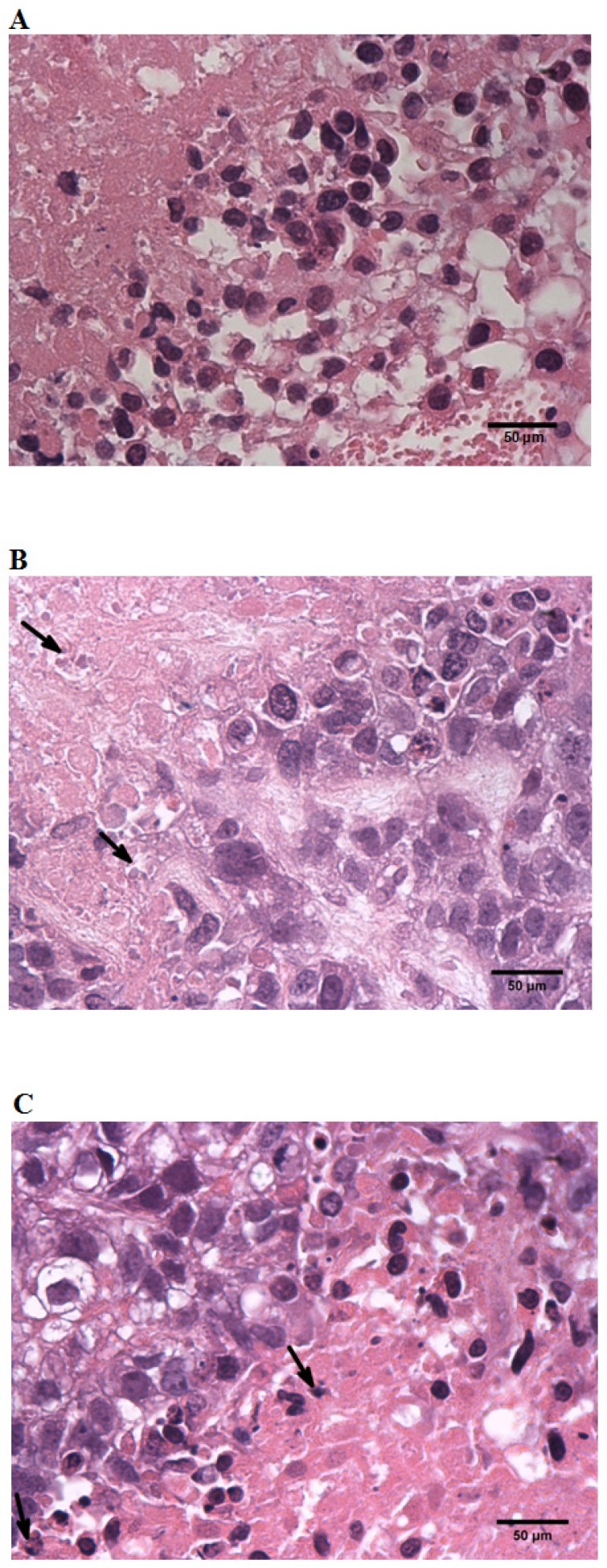
Figure 5.
Histology of SGC-7901 tumor from SCID mice. A, A section from tumor-cells of animals treated with negative control sera (N.C.) ×40. B, A section from tumor-cells of animals treated with standard high-dose chemotherapeutic combination protocols (H.C.) ×40. C, A section from tumor-cells of animals treated with combined-high dosage (H) group protocols ×40. All tumors were formalin fixed, paraffin embedded, and stained with hematoxylin and eosin. All assessments were made “blind” by an independent pathologist.

