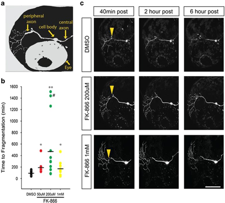
Figure 6.
FK866 delays Wallerian degeneration in vivo. (a) Schematic of larval zebrafish head indicating position of trigeminal neurons and axons, relative to the fish eye. (b) Time to beginning of fragmentation following laser axotomy in larvae pretreated for 2 h with vehicle (1% DMSO) (n=25), 50 μM (n=9), 200 μM (n=11) or 1 mM (n=10) FK866. Each circle represents one experiment; horizontal bar denotes average degeneration time (# indicates data from axon still intact >24 h, *P<0.05; **P<0.001). (c) Confocal images of trigeminal neurons postaxotomy labeled with DsRed-Express and treated with 1% DMSO or FK866. Arrowheads point to the site of axotomy. Scale bar, 100 μm

