Figure 4.
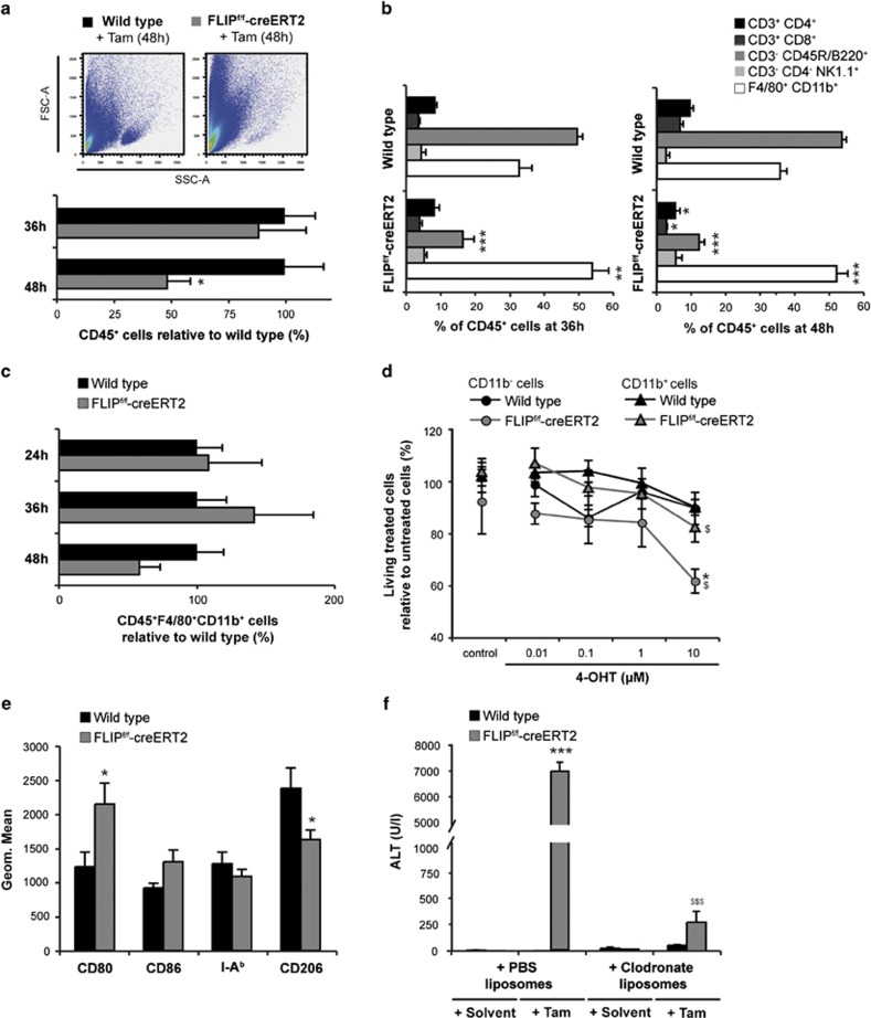
Loss of cFLIP induces apoptosis of intrahepatic T and B lymphocytes and activation of macrophages. (a) Living intrahepatic CD45+ cells were quantified by flow cytometry. Numbers are expressed relative to tamoxifen-treated wt mice. A representative forward (FSC) and side scatter (SSC) plot is shown. (b) Intrahepatic leukocyte subsets were quantified by gating on CD45+CD3+CD4+ and CD45+CD3+CD8+ for T lymphocytes, CD45+CD3−B220+ for B lymphocytes, CD45+CD3−CD4−NK1.1+ for NK cells and CD45+F4/80+CD11b+ for macrophages and quantitated relative to CD45+ cell numbers. (c) Intrahepatic macrophages were quantified by gating on living CD45+F4/80+CD11b+ cells. Numbers are expressed relative to tamoxifen-treated wt mice. (d) CD11b− and CD11b+ cells were isolated from spleen of naive wt and FLIPf/f-creERT2 mice and stimulated for 48h with different concentrations of 4-hydroxytamoxifen (4-OHT) as indicated or ethanol as a control. Cellular viability after deletion of cFLIP was quantified by Nicoletti assay. (e) Macrophages were characterized by M1/M2 markers. (f) Depletion of macrophages was achieved by clodronate-liposomes 48 h before tamoxifen. ALT was determined at 48 h. Data represent mean±S.E.M. (a–f: n=5–14 mice/group), P-values for wt versus FLIPf/f-creERT2: *P<0.05, **P<0.01, ***P<0.001 and (d) for FLIPf/f-creERT2 cells±zVAD and (f) for FLIPf/f-creERT2 mice±clodronate liposomes: $P<0.05, $$P<0.01, $$$P<0.001

