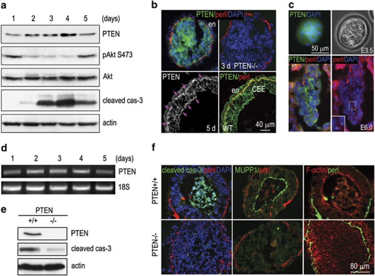
Figure 1.
Ablation of PTEN inhibits apoptosis, cavitation and epiblast polarization. (a) Normal EBs cultured for 1–5 days were analyzed by immunoblotting for PTEN, phospho-Akt Ser 473 (pAkt S473), Akt and cleaved caspase-3 (cas-3). Actin serves as a loading control. Expression of PTEN was increased in 2-, 3- and 4-day EBs, in parallel with reduced Akt phosphorylation and increased cas-3 activation. (b) Immunostaining showed that PTEN was highly expressed in the centrally located cells in 3-day EBs and was redistributed to the apical and the basal side of the epiblast epithelium in 5-day EBs (arrows). PTEN−/− EBs serve as a negative control for PTEN immunostaining. (c) E3.5 (early blastocyst stage) and E6.0 embryos were immunostained for PTEN and perlecan. PTEN was enriched in the centrally located cells. (d) Reverse transcription-PCR (RT-PCR) showed that PTEN mRNA levels were increased in 2-, 3- and 4-day EBs. (e) Three-day PTEN+/+ and PTEN−/− EBs were analyzed by immunoblotting for PTEN and cleaved cas-3. Ablation of PTEN inhibited cas-3 activation. (f) Five-day EBs were immunostained for cleaved cas-3 and the apical polarity marker MUPP1. Basement membrane was identified by perlecan immunofluorescence. F-actin was stained with rhodamine-phalloidin. Nuclei were counterstained with DAPI (4',6-diamidino-2-phenylindole). Ablation of PTEN inhibited apoptosis of the core cells and blocked cavitation. The epiblast cells of the mutant EBs failed to form a polarized apical domain

