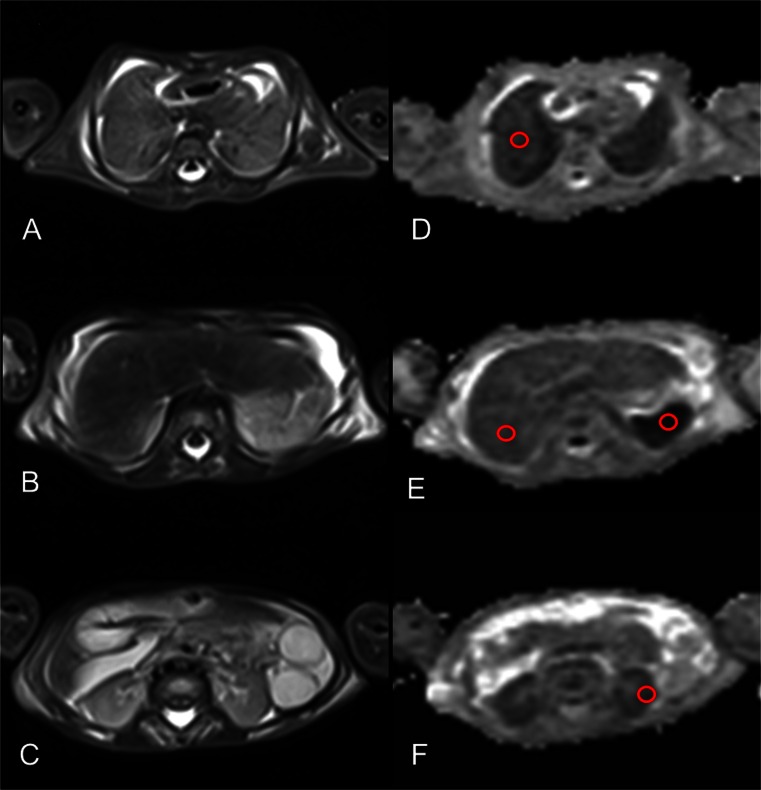
Fig. 2.
Thoraco-abdominal post-mortem magnetic resonance (PMMR) diffusion-weighted imaging (DWI). Axial T2-weighted MR imaging (left; a - c) and apparent diffusion coefficient (ADC) maps from DWI sequences (right; d - f) in a 36-week gestation foetus that underwent PMMR for a congenital brain malformation (not shown). Axial slices illustrate the lungs (a), liver and spleen (b) and kidneys (c), with corresponding regions of interest drawn on ADC maps over the lungs (d), liver and spleen (e), renal cortex (f) and muscle (not shown)

