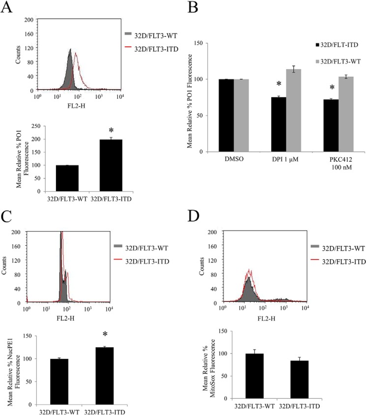
FIGURE 2.
32D stably transfected with FLT3-ITD have higher levels of endogenous H2O2 outside and inside the nucleus than 32Ds transfected with FLT3-WT. The inhibition of FLT3 receptor with PKC412 or NOX protein with DPI, causes a decrease in H2O2 in 32D/FLT3-ITD cells but not in 32D/FLT3-WT cells. The cells were IL-3 starved overnight, followed by ROS probe staining for 1 h before FACS reading. Flow cytometric PO1 analysis of total H2O2 (A and B), NucPE1 analysis of nuclear ROS (C), and MitoSOX analysis of mitochondrial ROS (D) in 32D expressing FLT3-WT or FLT3-ITD. In B, the 32D cells were treated for 24 h with DPI or PKC412 at the indicated concentrations. DMSO, dimethyl sulfoxide. The bar charts show the relative mean fluorescence of treated cells expressed as a percentage of control. The mean is representative of three independent experiments. The asterisk indicates statistically significant difference (p < 0.05) as analyzed by Student's t test. The error bars represent ± S.D.

